
Proton Therapy - Technology Transfer
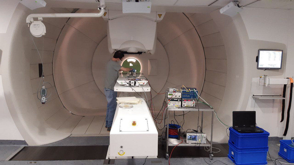
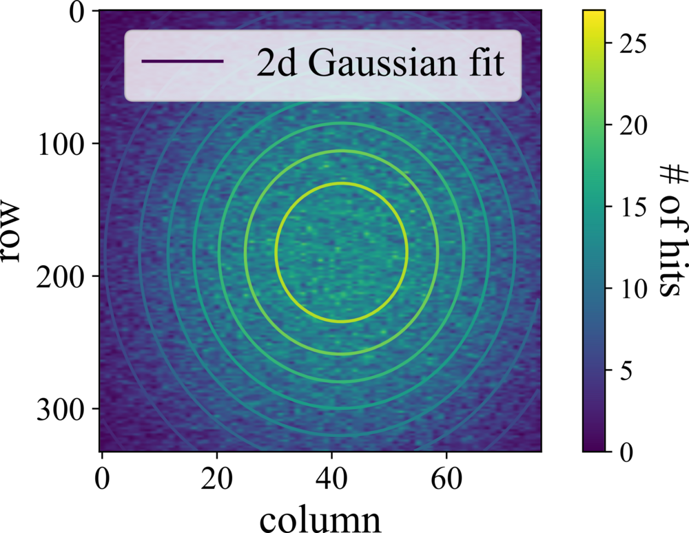
Proton therapy has proven to be very effective in the treatment of various types of tumors, as it allows to deposit the prescribed dose in a well-defined area. Pencil beam scanning (PBS) has quickly become the preferred method of dose delivery. A small-diameter (5-10 mm) beam is scanned in energy slices across the target volume, ideally irradiating the entire tumor while the surrounding healthy tissue to the sides and behind the tumor receives virtually no dose. To ensure patient safety and treatment efficacy, a task group of the American Association of Physicists in Medicine has published 224 comprehensive quality assurance guidelines for proton therapy centers, which are divided into daily, monthly, and annual tasks. Daily quality assurance checks include verifying the spot shape, dose consistency, and pencil beam field coverage. Our group is investigating the applicability of silicon pixel detectors, developed for high energy physics, for these daily quality assurance measurements. The detectors offer extreme radiation hardness, excellent spatial resolution and high readout speed, making them very well suited for this application.

To be able to control the positioning of patients in real time during irradiation, we are working on using the same pixel detectors to perform radiography with protons. For this purpose, the energy of the protons is briefly increased during the treatment to such an extent that they no longer stop in the tissue, but reach the detectors mounted behind the patients. A measurement of the number and energy of protons allows to distinguish between different types of tissues and thus to record an image of the patient. Modern image reconstruction algorithms are used to identify structures inside the patient. The range of protons in the patient is subject to several uncertainties that potentially limit the usefulness of proton therapy near organs at risk. One of such contributions is the uncertainty of the mass stopping power of the tissue, which is obtained using a calibration from the electron density measured in the planning CT. To reduce the uncertainty due to this calibration, we are investigating direct measurements of the mass stopping power of protons in tissue. The pixel detectors allow to measure the energy of the protons hitting them. If the trajectory of individual protons through the patient can be determined, the distribution of the mass stopping power can be determined via the energy the protons deposited along this trajectory.
Funded by: